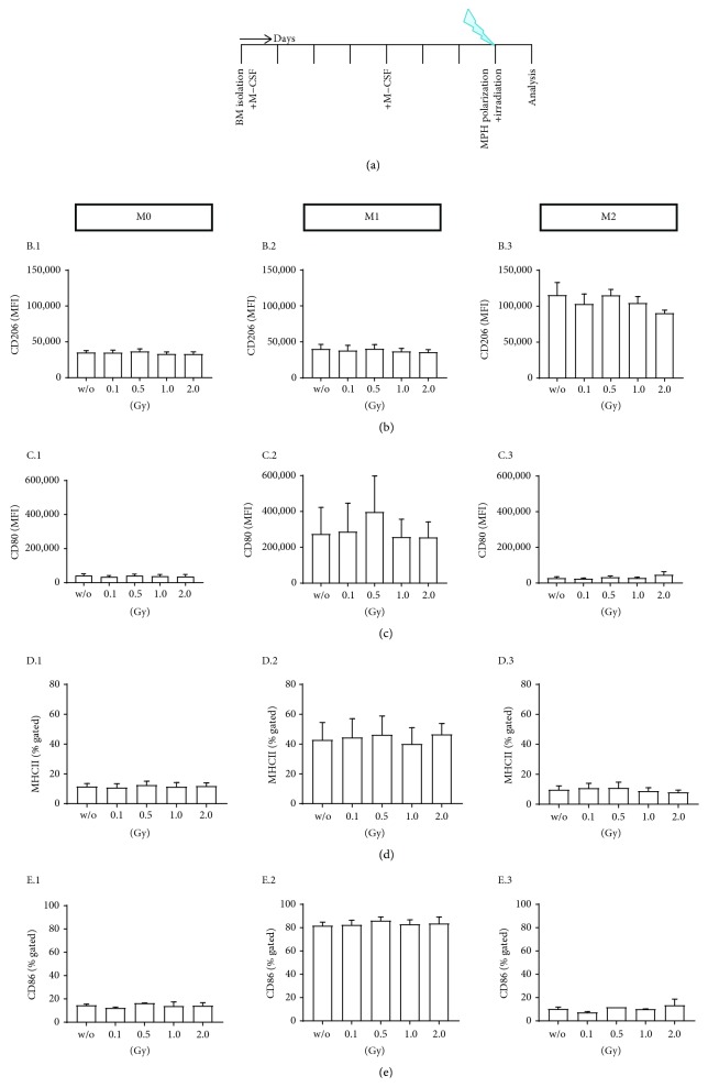
Figure 2.
Low-dose radiotherapy has no influence on macrophage polarization. hTNF-α tg bone marrow was isolated and differentiated into M0 macrophages according to the experimental set-up shown in (a). 24 h prior to the characterization of the macrophage phenotypes, cells were treated with three different polarization cocktails (M0: 5 ng/ml M-CSF; M1: 4 ng/ml M-CSF, 20 ng/ml IFN-γ, and 20 ng/ml LPS; M2: 5 ng/ml M-CSF and 20 ng/ml IL-4). 2 h after this stimulation, cells were irradiated with the indicated dose. Macrophages showed characteristic surface marker expression after polarization such as CD206 ((b) B.1–B.3) for M2 and CD80 ((c) C.1–C.3), MHCII ((d) D.1–D.3), and CD86 ((e) E.1–E.3) for M1. No significant differences of the surface marker expression were found in dependence of irradiation and irradiation doses. Depicted is data from three independent experiments. Data is presented as mean mean ± SEM.

