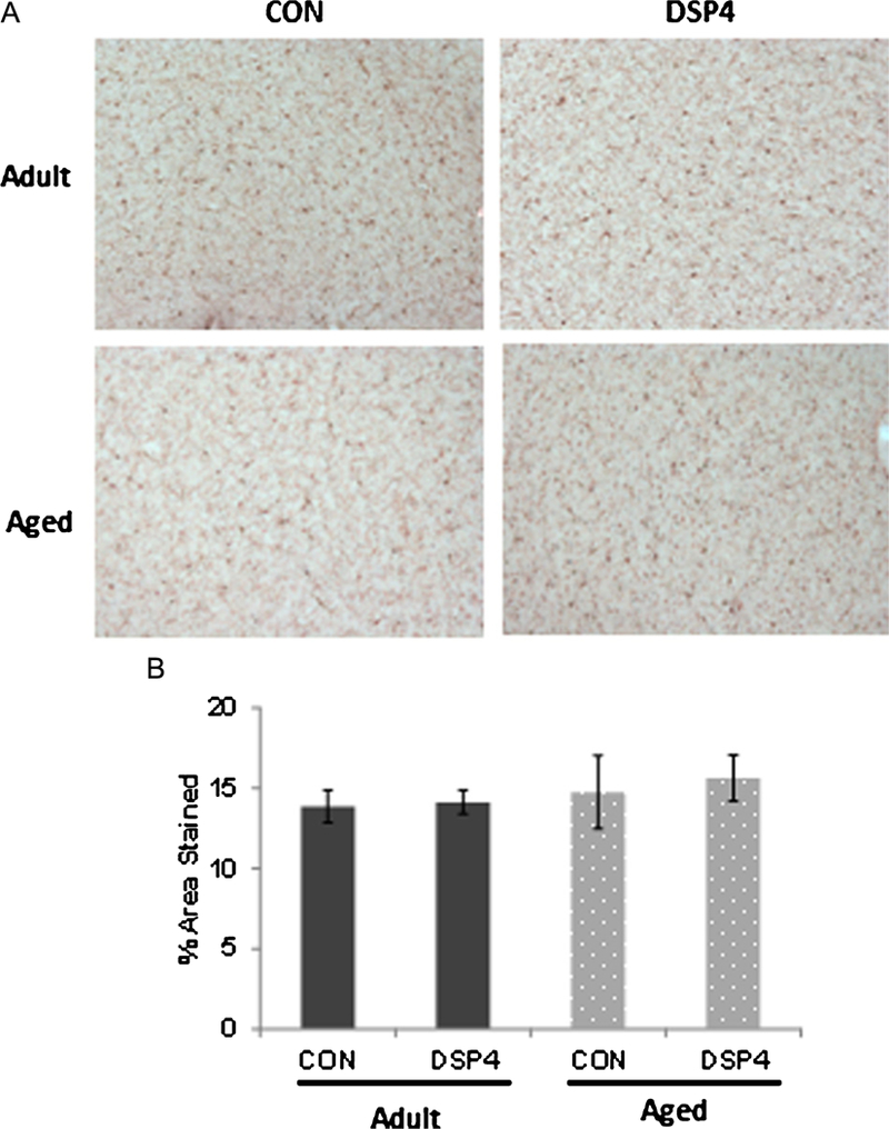
Fig. 6.

Microglia distribution in cortex. A) Microglia were widely distributed throughout the brain; staining pattern was similar across adult and aged monkeys regardless of treatment group. B) Semi-quantitative determination of % area stained in serial sections from frontal cortex (p > 0.05). DSP4: nadult = 6 and naged = 3 Control: nadult = 5 and naged = 3.
