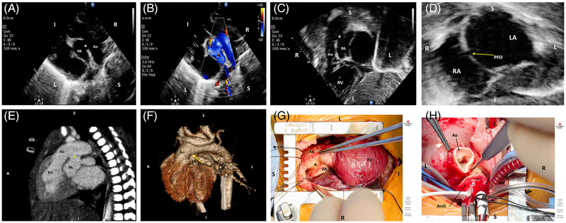
FIGURE 1.
A, Two-dimensional transthoracic echocardiographic parasternal long-axis view demonstrating the aortopulmonary window (asterisk) in relation to a rightwards aorta (Ao) and leftwards pulmonary artery (PA). B, Parasternal long-axis color Doppler view revealing shunting at the aortopulmonary window (asterisk), shunting systemic-to-pulmonary in this view. C, Two-dimensional transthoracic echocardiographic subcostal view showing the rightwards aorta (Ao) arising off the anterior right ventricle (RV) and dropout of the great vessel wall at the level of the aortopulmonary window (asterisk). D, Two-dimensional transthoracic echocardiographic subcostal view at the level of the atrial septum demonstrating a patent foramen ovale (arrow) with an otherwise intact atrial septum, with left atrium (LA) on the left and right atrium (RA) on the right. E, Cardiac computerized tomographic sagittal view demonstrating transposition anatomy with the aorta (Ao) anterior and arising off a rightwards RV, a posterior PA, and the aortopulmonary window (asterisk) measuring 10 mm in diameter and originating 10 mm distal to the aortic valve leaflets. F, Cardiac computerized tomographic 3D reconstruction demonstrating transposition anatomy with the area of the aortopulmonary window (arrow). G, Intra-operative findings of a type B aortopulmonary window (asterisk) between the anterior aorta (Ao) and posterior PA. The RV is noted to be the anterior ventricle. H, A trans-aortic approach was undertaken for repair of the aortopulmonary window. The transected aorta (Ao) and proximal transverse arch can be seen here in the arterial switch component of the operation

