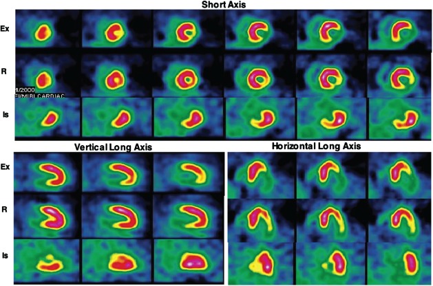
Figure 2.

Representative exercise (Ex) and rest (R) technetium‐99 m‐sestamibi and exercise 18 F‐fluorodeoxyglucose (18FDG) images of a patient with angina in the short axis, and vertical and horizontal long axes. This patient had no history of myocardial infarction. A large area of partially reversible perfusion abnormality involving the inferior and lateral walls is seen on the perfusion images. Intense 18FDG uptake is seen in the areas corresponding to the perfusion abnormalities. On coronary angiography, 100% occlusion of the right coronary artery and a 50% narrowing of the left anterior descending coronary artery were observed. Reproduced with permission from Jain and He.34
