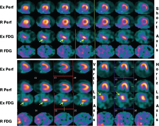
Figure 4.

This 49‐year‐old male with exertional angina underwent exercise (Ex) and rest 99mTc‐sestamibi (Perf) and 18 F‐fluorodeoxyglucose (FDG) imaging 24 hours apart. Representative short axis, and vertical (Verti) and horizontal (Horiz) long (Lg) axes slices of the heart are shown. Reversible perfusion abnormality in the posterior septum and inferior walls is observed on this study. Intense FDG uptake on the exercise images (yellow arrows) is seen in the corresponding segments. No FDG uptake is seen in the heart on rest images. This patient was found to have 85% narrowing of the right coronary artery. Reproduced with permission from Dou et al.40
