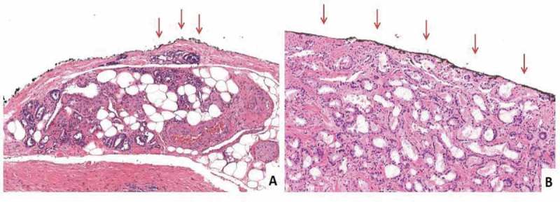
Figure 1.

Histopathological images of focal and non-focal PSMs. The arrows indicate the PSM site. (A) The inked margin has neoplastic cells with a length ≤1 mm, i.e. focal PSM (H&E, ×10). (B) The inked margin has neoplastic cells with a length >1 mm, i.e. non-focal (in the present case, the entire inked margin is involved. H&E, ×10).
