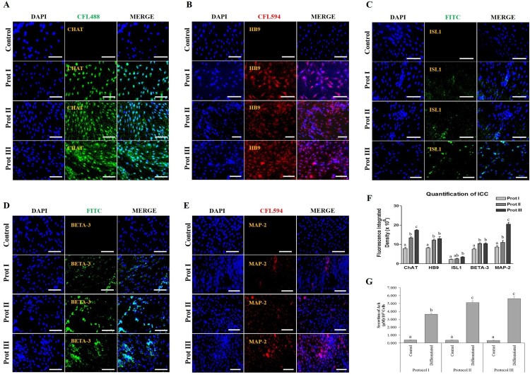
Figure 6.
Immunocytochemical (ICC) analysis and the functional properties of in vitro differentiated cholinergic neurons. (A-E) Differentiated cells under all the three protocols strongly expressed cholinergic-specific proteins such as ChAT, HB9, ISL1, BETA-3, and MAP-2, whereas the same proteins were not expressed in undifferentiated control DPSCs (Scale bar = 100 µm). (F) Quantification analysis of ICC revealed that the differentiated cells under protocol III showed higher expression of cholinergic marker proteins compared to the other two protocols. (G) Quantification of acetylcholine (Ach) by ELISA. The level of Ach was analysed in the culture media using a biochemical fluorescent assay, indicating that all the differentiated cholinergic neurons could synthesize Ach. However, protocol III showed the highest Ach secretion. Data represent mean ± SEM from three independent experiments. Significant differences are denoted using different letters when p < 0.05.

