Figure 2.
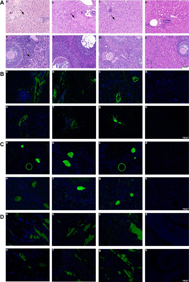
Histopathology and immunofluorescence of pregnant rabbits in group A, B, C and D. Liver and ovary histopathology. (a-c) HE staining showed hepatocyte degeneration and necrosis, collagen fibre hyperplasia and fibrosis, and infiltration of inflammatory cells in liver sections of group D, E and F rabbits; (d) liver section of group G showed no visible pathological changes; (e) mild inflammatory cells’ inflitration in ovary section of group A; (f-h) no visible pathological signs of HEV infection in ovary sections of group B-D rabbits. (B) Immunofluorescence staining of HEV ORF2 and ORF3 in liver. (a-c, e-g) Positive signals of HEV ORF2 and ORF3 respectively in liver sections of rabbits in group A-C; (d, h) no positive signals of HEV ORF2 and ORF3 were observed in liver section of rabbits in group D. (C) Immunofluorescence staining of HEV ORF2 and ORF3 in ovary. (a-c, e-g) Positive signals of HEV ORF2 and ORF3 respectively in ovary sections of rabbits in group A-C; (d, h) no positive signals of HEV ORF2 and ORF3 were observed in ovary section of rabbits in group D. (D) Immunofluorescence staining of HEV ORF2 and ORF3 in placenta. (a-c, e-g) Positive signals of HEV ORF2 and ORF3 respectively in placenta sections of rabbits in group A-C; (d, h) no positive signals of HEV ORF2 and ORF3 were observed in placenta section of rabbits in group D. Original magnification,×200.

