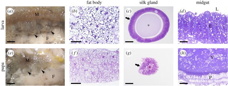
Figure 1.
Remodelling of silkworm organs during metamorphosis. (a,e) Stereomicroscopic images showing the internal anatomy of Bombyx mori; (b,f) cross-sections of the fat body; (c,g) cross-sections of the silk gland; (d,h) cross-sections of the midgut. E, larval midgut epithelium; F, fat body; Y, yellow body; L, midgut lumen; M, midgut; P, pupal midgut epithelium; arrowheads, silk glands; arrows, silk gland epithelium; asterisk, silk gland lumen. Bars: 25 mm (a,e); 50 µm (b,f); 200 µm (c); 100 µm (d,g,h). (Online version in colour.)

