
FIGURE 2:
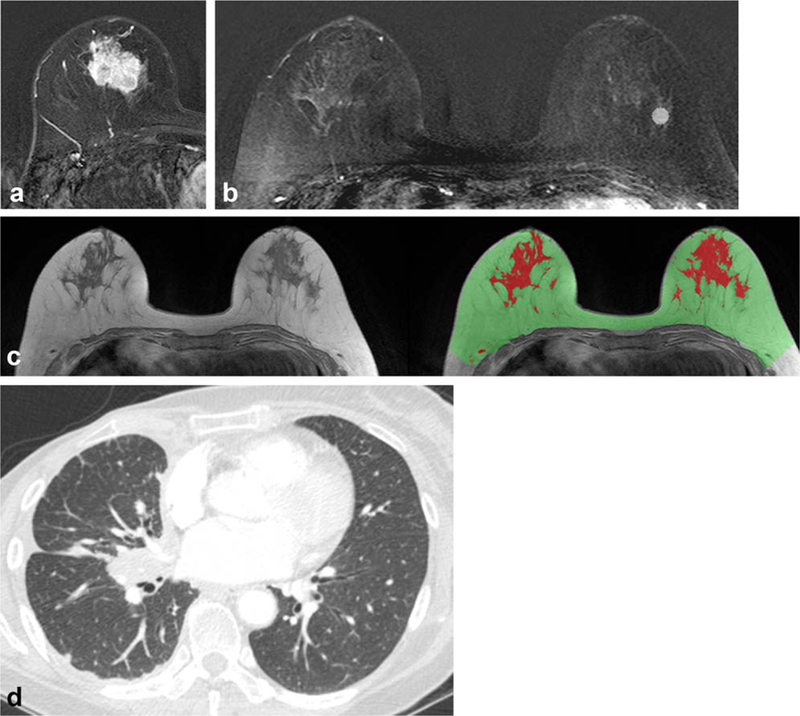
A 57-year-old woman with invasive ductal carcinoma in the right breast. (A) Axial T1-weighted contrast-enhanced early subtraction MR image shows a 25-mm size invasive breast cancer at the right breast, with a high Ki-67 level of 30%. (B) Axial T1-weighted contrast-enhanced early subtraction MR image shows the location of one of the three ROIs used for ROI-based quantitative BPE analysis. (C) T1-weighted precontrast MR imaging without fat suppression is shown with the breast parenchymal mask (area in green and red) and fibroglandular tissue segmentation (area in red). (D) Patient underwent right mastectomy, adjuvant chemotherapy, and endocrine therapy but developed lung metastasis 49.4 months after diagnosis. Chest CT image shows right pleural and interlobar lymph node metastasis.