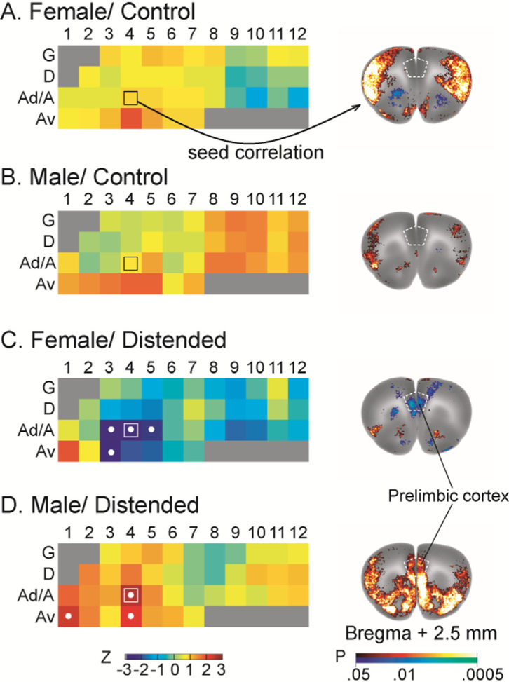
Fig. 7.
Sex and CRD-related differences in insular functional connectivity with the prelimbic area of the medial prefrontal cortex. The strength of prelimbic functional connectivity with individual insular region of interest is color coded in insular flat maps (left column, with coding of insular regions as in Fig. 1). Regions showing statistically significant correlations are marked with white dots. Distension induced negative functional connectivity between the anterior/mid insular cortex and the prelimbic cortex in females, but positive connectivity in males. Representative seed correlation results using Ad4 (marked with □ on the flat map) as the seed further showed this striking sex difference (right column). Color-coded overlay over the template brain at bregma +2.5 mm (right column) shows brain areas that are significantly correlated with the insular seed (P < 0.05 for clusters of > 100 contiguous, significant voxels).

