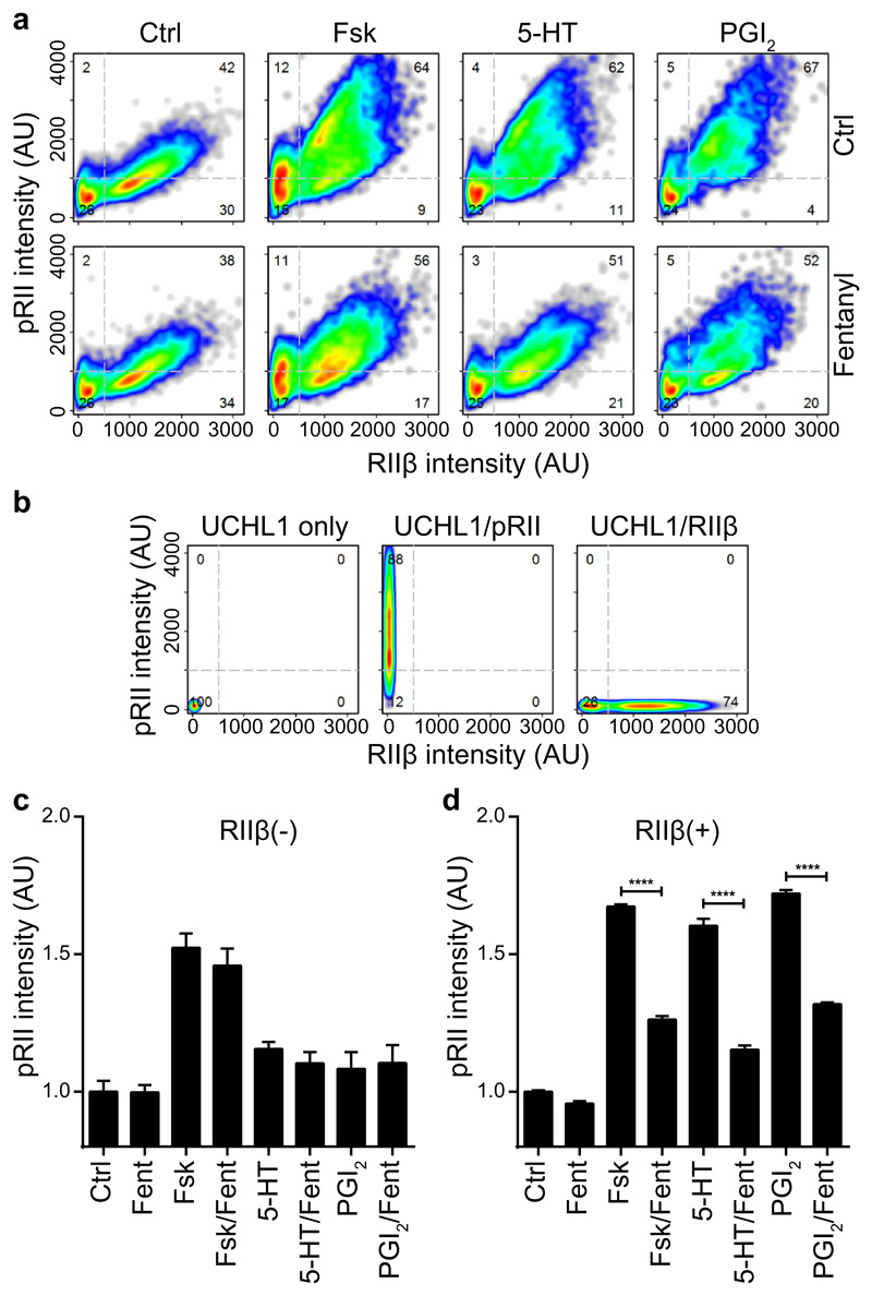
Fig. 2. Opioids inhibit pRII-increases selectively in RIIβ(+) sensory neurons of rats.
(A) Cell density plots showing single cell data of pRII/RIIβ-labeled rat sensory neurons stimulated with Fsk (2 µM), 5-HT (0.2 µM), or PGI2 (1 µM) in the absence (top) or presence (bottom) of fentanyl (2 µM). Dashed lines indicate gating thresholds to discriminate between RIIβ(-) and RIIβ(+) neurons with the numbers indicating the relative percentage of cells in the respective quadrant. Combined data of n = 4 experiments with a total of >8000 neurons/condition. (B) Compensation controls showing removal of spill-over between fluorescence channels. The graphs show the pRII versus the RIIβ intensity in control samples stained with UCHL1 alone (left), UCHL1 and pRII (middle), or UCHL1 and RIIβ (right) after compensation has been applied. (C) Normalized mean pRII intensities in RIIβ(-) (left) and RIIβ(+) (right) neurons. Values are means ± SEM; n = 4 independent experiments; >8000 neurons/condition; two-way ANOVA with Bonferroni’s test; *P<0.05; **P<0.01; ***P<0.001; ****P<0.0001.

