
Figure 5.
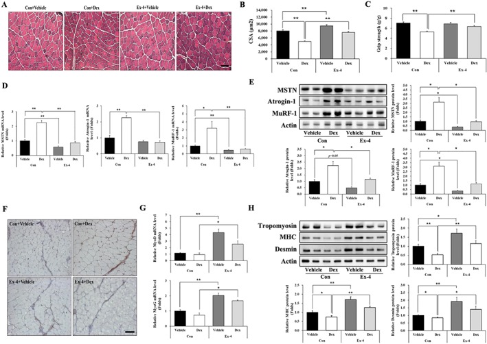
Ex‐4 recovers muscle strength in Dex‐induced muscle atrophy mice. (A) Frozen serial transverse cryosections (7 μm) from TA muscle tissue were stained with H&E and examined under a microscope (magnification ×200). (B) The cross‐sectional area (CSA) of muscle fibre was measured using Image J program. (C) Muscle function was assessed using grip strength measurement. The grip strength was normalized to the final body weight (g). (D–E) The mRNA level of muscle atrophic factors (MSTN, atrogin‐1, and MuRF‐1) (D) and protein (E) levels were assessed using RT‐QPCR and western blotting in TA muscle tissue. (F) The TA muscle tissue from mice administered with Dex was immunostained with anti‐MSTN antibody. The image was taken under a confocal microscope (magnification ×200). Brown colour indicates MSTN expression. (G) The mRNA levels of myogenic factors (MyoD and MyoG) were assessed using RT‐QPCR in TA muscle tissue. (H) The protein levels of myofibrillar proteins (tropomyosin, MHC, and desmin) were measured using western blot. All values are expressed as the mean ± standard error. Significant differences are indicated as **P < 0.01, *P < 0.05 compared with Con + vehicle or Con + Dex, n = 5/group. Con, control; Dex, dexamethasone; Ex‐4, exendin‐4.