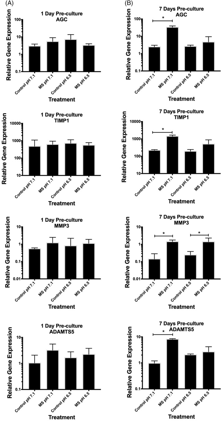
Figure 2.

Nucleus pulposus (NP) cells (n = 3) were encapsulated (2 × 10⁶ cells/mL) in 2% agarose gel and cultured for (A) 1 day or (B) 7 days in standard Dulbecco's modified Eagle's medium (DMEM) medium. Encapsulated cells were then cultured for a further 24 hours in DMEM at pH 7.1 or 6.5 (to mimic the nondegenerate or degenerate intervertebral disc [IVD] microenvironment, respectively) and then mechanically compressed with 0.004 MPa at 1.0 Hz for 1 hour. Uncompressed encapsulated cells served as the control. Gene expression is presented using the 2‐dCt method34 relative to MRPL19 and EIF2B1. (A) Compression of encapsulated cells following 1 day of preculture in standard DMEM resulted in no change to the expression of any genes assessed, at either pH 7.1 or 6.5. (B) However, both anabolic/anti‐catabolic genes (aggrecan [AGC] and tissue inhibitor of metalloproteinases‐1 [TIMP1]) increased in NP cells compressed at pH 7.4, but not at pH 6.5, following 7 days of preculture in standard medium. The catabolic genes (matrix metalloproteinase‐3 [MMP3] and a disintegrin and metalloproteinase with thrombospondin motifs‐5 [ADAMTS5]) both increased in NP cells precultured for 7 days in standard medium and then compressed at pH 7.1. Interestingly, MMP3 gene expression was also increased in NP cells compressed at pH 6.5. The Mann‐Whitney U test was used to test for significance between control and compressed treatments, with P ≤ .05 considered significant and indicated by “*”
