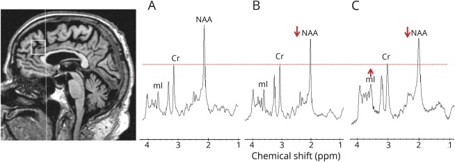
Figure 1. Voxel location and representative 1H MRS from all participants.
Medial frontal lobe voxel is placed on a midsagittal 3D T1-weighted image (left). Voxel landmarks include the following: (1) the posterior edge of the frontal lobe voxel is in alignment with the posterior border of the genu of the corpus callosum; and (2) the posterior lower corner is at the superior border of the corpus callosum. Examples of proton magnetic resonance spectra (1H MRS) in (A) a noncarrier, (B) an asymptomatic microtubule-associated protein tau (MAPT) mutation carrier, and (C) a patient with behavioral variant frontotemporal dementia (bvFTD) with MAPT mutation. Spectra are scaled to the creatine (Cr) peak as indicated with the dotted red line. The N-acetylaspartate (NAA) peak is decreased in (B) the asymptomatic MAPT mutation carrier and (C) the patient with bvFTD. The myo-inositol (mI) peak is elevated only in (C) the patient with bvFTD.

