. 2019 Aug 27;8:e46134. doi: 10.7554/eLife.46134

Figure 1. Design and validation of FLAgs as activators of the Wnt-βcatenin pathway.

(A) Surface plasmon resonance (SPR) binding kinetics of FP Fab. Kinetics were derived from curves for soluble FP Fab interacting with immobilized FZD CRD, and ‘x’ indicates no detectable binding. (B) Anti-LRP6 Ab inhibitory activity. Inhibition of WNT1 or WNT3A signaling by indicated LRP6 Abs in the diabody-Fc format was assessed using stimulation with purified WNT3A or upon WNT1 cDNA transfection. (C) Molecular architecture of tetravalent FLAgs. (D) Activation of βcatenin signaling by FLAgs. Dose response curves are shown for the activation of a LEF/TCF reporter gene (y-axis) in HEK293T cells by serial dilutions of pan-specific FLAg proteins (FP+P-L61+1, FP+P-L63+3 and FP+P-L61+3) (x-axis). Error bars indicate SEM, n = 3. (E) Levels of βcatenin protein in RKO cells after 30 min treatment with the indicated concentrations of pan-FLAg (FP+P-L61+3). Representative blot of three replicates. (F) Time course of βcatenin and phosphorylated Disheveled-2 (p-DVL2) protein levels in RKO cells treated with 10 nM pan-FLAg (FP+P-L61+3). Representative blot of three replicates.

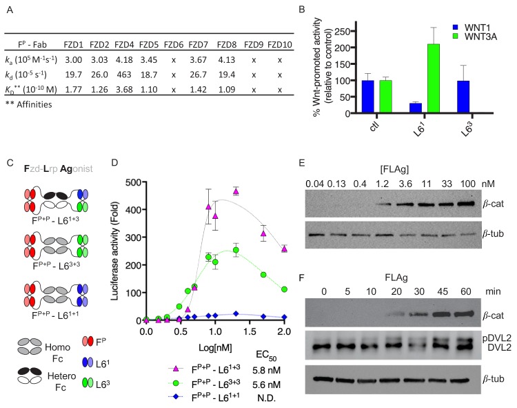
Figure 1—source data 1. Source data for Figure 1B.
DOI: 10.7554/eLife.46134.004
Figure 1—source data 2. Source data for Figure 1B.
DOI: 10.7554/eLife.46134.005

Figure 1.

Figure 1—figure supplement 1. Development of Synthetic Modular Tetravalent FZD.

Figure 1—figure supplement 1.

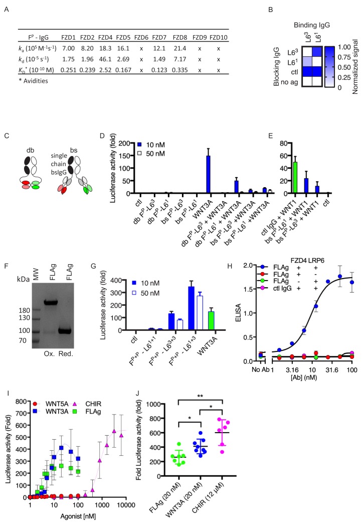
(A) SPR binding kinetics of FP IgG. 'x’ indicates no detectable binding. (B) Epitope binning for L61 and L63 IgG by competitive blocking measured using BLI. The normalized signal is shown for the binding of L61 or L63 (x-axis) to immobilized LRP6-Fc pre-blocked with the indicated Ab (y-axis). (C) Schematic representation of bispecific diabody-Fc (db) and single-chain bispecific IgG (bs) modalities recognizing FZD and LRP6 receptors. (D–E) Effects of bispecific IgGs and diabody-Fcs on Wnt-βcatenin signaling, as measured using HEK29T cells stably expressing a LEF/TCF reporter and with or without WNT1 transient transfection or treatment with WNT3A (0.5 μg/ml). (F) Coomassie-stained SDS-PAGE analysis of purified FLAg FP+P-L61+3 under oxidizing (Ox) or reducing (red) conditions. (G) βcatenin signaling stimulated by FLAg or WNT3A conditioned media, measured using LEF/TCF reporter assay in HEK293T cells. (H) Co-binding of FLAg FP+P-L61+3 to absorbed LRP6-Fc (LRP6) and soluble concentrations of FZD4-His (FZD4) by ELISA with anti-His-HRP detection. (I) Dose response analysis of βcatenin signaling activity stimulated with mouse WNT3A (R and D systems 1324-WN/CF), human/mouse WNT5A (R and D systems, 645-WN/CF) purified proteins, FLAg FP+P-L61+3 or CHIR99021. HEK293T cells stably expressing a LEF/TCF reporter were stimulated with the indicated doses for 16 hr. (J) The fold change of luciferase activity at maximal stimulation of Wnt reporter cells treated with 20 nM FLAg FP+P-L61+3 , 20 nM purified WNT3A, or 12 μM CHIR99021.