
Figure 5.
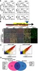
Aging and HF feeding alters the character of mesenchymal stem cells in sFAT (Ad-MSC). (A) Evaluation of Ad-MSC using a set of stem cell markers by Flow Cytometry. (B) Assessment of the differentiation capacity of Ad-MSCs into brown adipocytes (BA) using AdipoInducer Reagent, pioglitazone (1 µM) and CL316,243 (2 µM). The efficiency of BA induction was assessed by UCP1 IHC, mitochondrial (MitoGreen) and oil-red O staining. Pre-BAs from the classical BAT of 6-week-old male C57BL/6NCrSlc mice were used as positive control. (C) Scatterplot of gene expression profiles for Ad-MSCs from 24w/C and 12w/C mice (left panel) or from 24w/HF and 12w/HF mice (right panel). Green lines indicate the cut-offs for 2-fold up- and down-regulation. (D) Venn diagram of the down-regulated genes (with p<0.05 and >2-fold difference) in Ad-MSCs in the process of senescence (from younger to older mice). In the process of senescence, 2,190 genes in the control diet feeding groups and 2,302 genes in HF feeding groups were down-regulated. 798 genes were common to both categories. Several genes associated with browning in sFAT were included in each category, as shown in a schema.