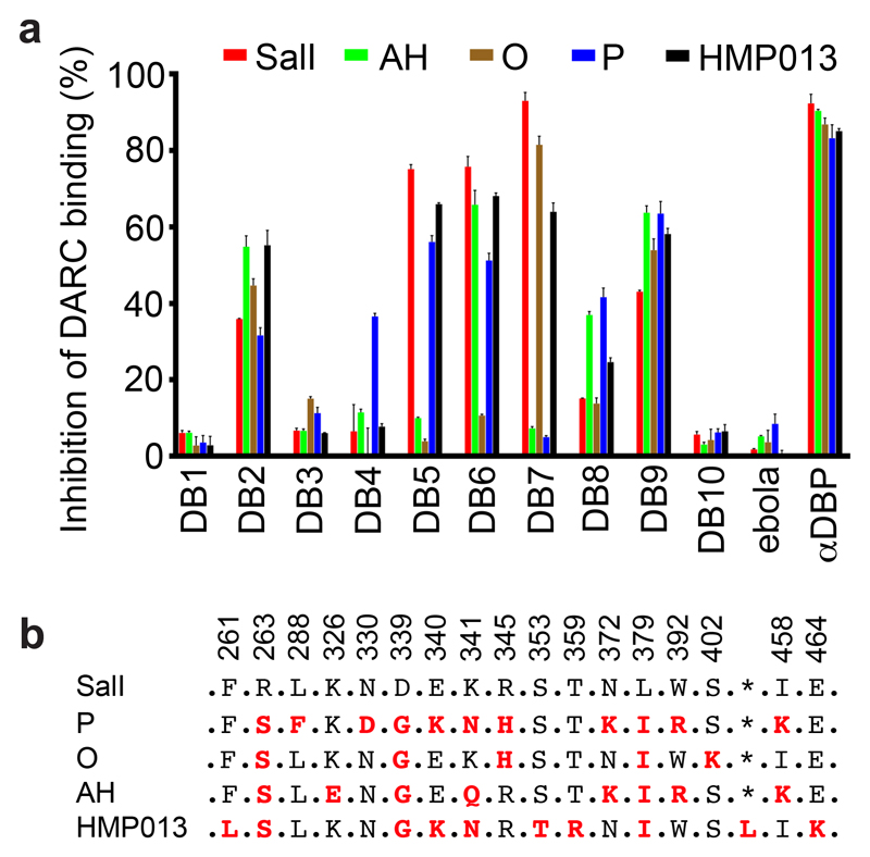
Figure 2. Inhibition of the binding of recombinant PvDBPII to DARC ectodomain.
(A) Assessment of the % binding of five naturally occurring variants of PvDBPII to the DARC ectodomain in vitro in the presence of 100 μg/mL concentration of each mAb (DB1-DB10). Individual titration curves are shown in Supplementary Figure 2. “αDBP” is polyclonal human anti-PvDBPII serum at 1:5 dilution while ‘ebola’ is an anti-Ebolavirus recombinant human IgG1 mAb included as a negative control. Data points represent the mean of three technical replicates, while the error bars represent the standard deviation. (B) Sequence polymorphisms of PvDBPII variants used in the assay. Numbering is according to the SalI reference sequence. Amino acid polymorphisms are indicated for the PvDBPII variants (P, O, AH and HMP013). Amino acids that are the same as the SalI reference sequence are black, those divergent from SalI are red and * indicates the absence of a leucine insertion between V429 and P430 in HMP013.

