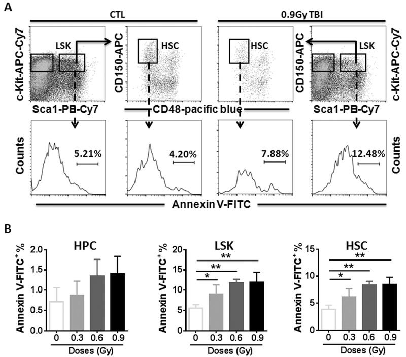
Fig. 3.
28Si TBI increases cellular apoptosis in HSCs. Lin− cells were isolated from control (CTL) and irradiated (TBI) mice two weeks after 28Si TBI as described. (A). Representative analysis of apoptosis by flow cytometry using Annexin V staining in BM HPCs, LSK cells and HSCs from control and irradiated mice. The numbers presented in the histograms are percentages of Annexin V-FITC positive cells in the indicated populations from a representative experiment. (B) The percentages of Annexin V positive cells in BM HPCs, LSK cells and HSCs after TBI are presented as mean ± SD (n = 5). * p < 0.05, ** p < 0.01 TBI vs. CTL.

