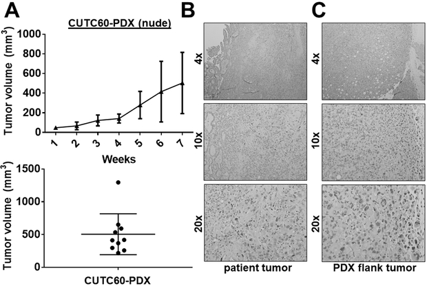
Figure 5. Generation of the CUTC60 patient-derived xenograft (PDX) model.
Tumor tissue (3 mm3) from patient 60 was directly implanted into the flanks of NSG mice by trocar injection. Tumors were passaged into the flanks of nude mice at generation mF3. A) The growth of the CUTC60-PDX was measured weekly for 7 weeks. Final tumor volumes are the mean +/− SD of 10 tumors. Representative images of the patient tumor (B) and PDX flank tumor (C) are shown.

