Figure 3.
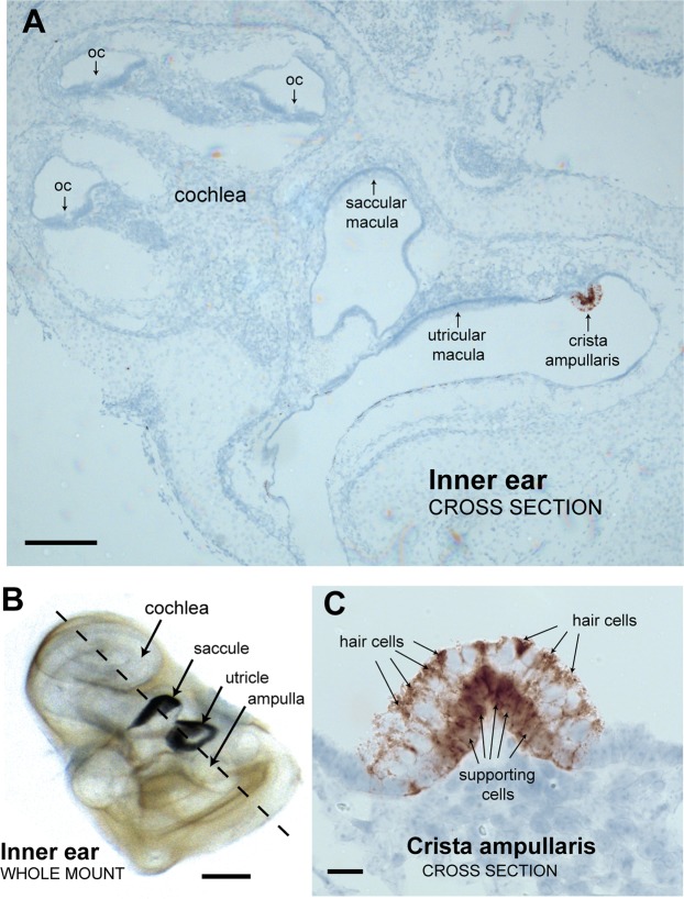
Zpld1 mRNA expression in the inner ear is primarily restricted to cells of the crista ampullaris. (A) In situ hybridization results showing a cross section of an inner ear from a newborn B6 mouse with areas of positive Zpld1 expression stained brown with a blue counter stain (Hematoxylin). Sensory epithelia with overlying acellular membranes are indicated by arrows, and include the organ of Corti (oc) in the cochlea, the saccular and utricular maculae, and the crista ampullaris. Zpld1 expression was detected only cells of the crista ampullaris. Scale bar, 200 μm. (B) A cleared, whole mount preparation of an inner ear from a newborn mouse positioned to match the orientation of the cross section shown in panel A. Otoconial crystals block microscope illumination from below, giving the saccular and utricular maculae a dark appearance. The dashed line indicates the corresponding plane of dissection. Scale bar, 500 μm. (C) Higher magnification of a crista ampullaris shows that Zpld1 appears to be expressed in both hair cells and supporting cells. Positive expressing cells at the crista surface were presumed to be hair cells, and positive expressing cells near the base were presumed to be supporting cells. Scale bar, 20 μm.

