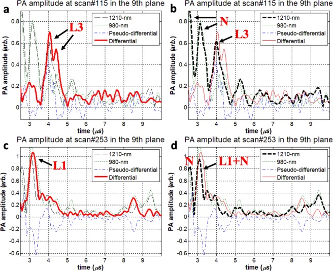
Figure 2.
Artificial plaque lump 3 (L3) signal was located very close to strong RF noise at scan #115 in the 9th imaging plane. From this measurement, (a) IV-DPAR amplitude and (b) LF single-ended 1210-nm PAR amplitude was highlighted. At scan #253 in the 9th imaging plane, the L1 signal was completely superposed with the local RF noise. From this measurement, (c) IV-DPAR amplitude and (d) LF single-ended 1210-nm PAR amplitude was highlighted. In both situations, IV-DPAR could successfully extract cholesterol signals from the background while the single-ended 1210-nm PAR mode was severely affected by the adjacent noise. N: RF + arterial wall noise.

