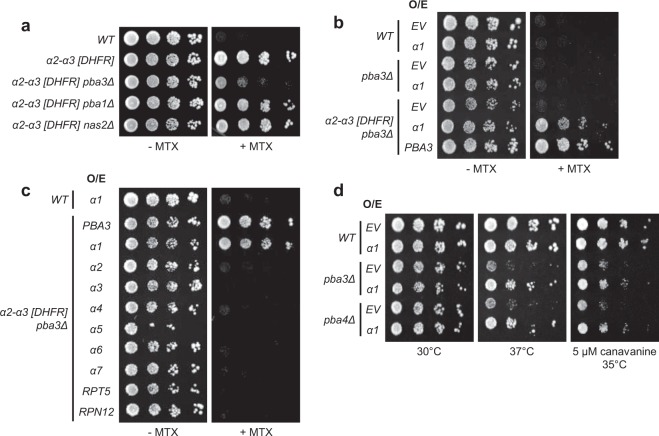
Figure 2.
A genome-wide screen identifies proteasome subunit α1 as an enhancer of α2-α3 juxtaposition in pba3Δ cells. (a) Equal numbers of cells from the indicated yeast strains were spotted in six-fold serial dilutions on synthetic complete plates lacking or containing MTX and incubated for three days at 30 °C. (b) Equal numbers of WT, pba3∆, or α2-α3 [DHFR] pba3∆ cells were transformed with empty vector (EV) or with high-copy plasmids encoding α1 or PBA3. Transformants were spotted in six-fold serial dilutions onto synthetic complete plates lacking or containing MTX and incubated for three days at 30 °C. (c) Equal numbers of WT or α2-α3 [DHFR] pba3∆ cells expressing the indicated proteins from high-copy plasmids were spotted in six-fold serial dilutions onto synthetic complete plates lacking or containing MTX and incubated for three days at 30 °C. (d) Equal numbers of cells from the indicated yeast strains expressing empty vector (EV) or α1 from a high-copy plasmid were spotted in six-fold serial dilutions on the indicated media and incubated as shown for three days.

