Figure 4.
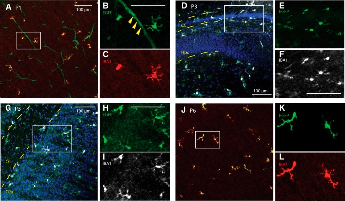
Tmem119-EGFP mice label microglia at early postnatal stages. Sagittal slices from P1, P3, and P6 mice were stained against IBA1. A–C, High-power confocal images of the somatosensory cortex show EGFP expression in IBA-positive cells and blood vessels at P1 (solid arrowheads in magnified panel). D–F, Confocal micrographs around the forceps major of corpus callosum (CC) and hippocampus (Hpc) show EGFP-labeled, IBA1-positive microglia at P3. G–I, Confocal micrographs of the striatum (CPu) and CC show EGFP labeling of IBA-positive cells at P3. J–L, Confocal micrographs of the P6 cortex reveal EGFP-expression in IBA-immunostained cells. Error bars, 100 μm.

