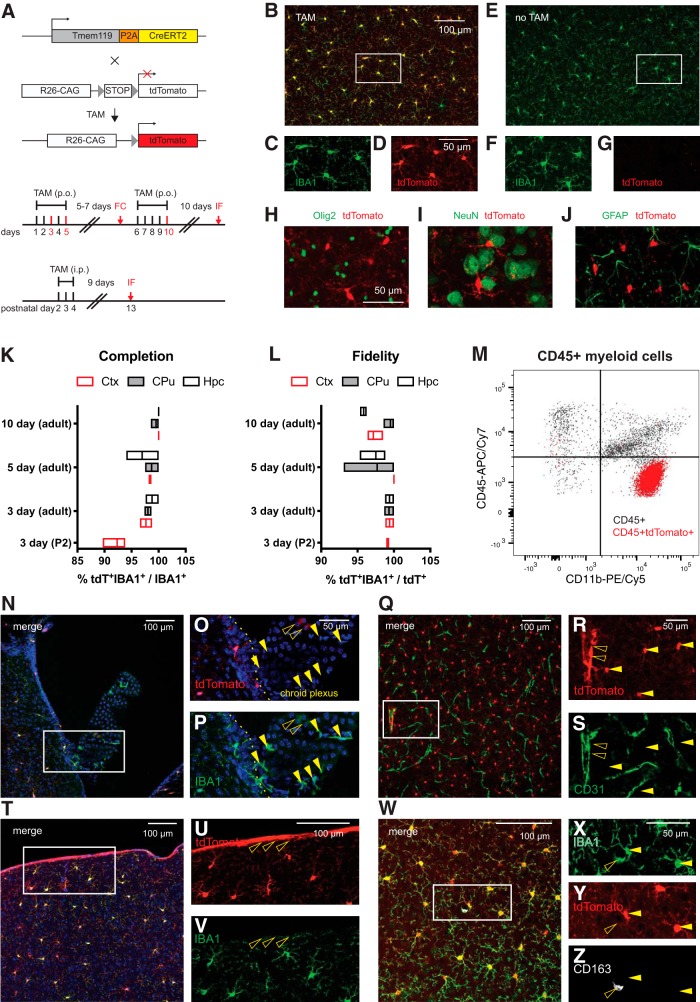
Figure 5.
Tmem119-CreERT2 mice effectively recombine a conditional allele in microglia. A, Tmem119-CreERT2 mice were crossed to Ai14(RCL-tdT)-D mice and tamoxifen (TAM) was administered to induce tdTomato expression. Different sets of adult mice received tamoxifen per os (p.o.) for 3, 5, or 10 d and a set of neonatal mice received TAM for 3 d intraperitoneally (i.p). The mice were killed for flow cytometry (FC) 7 d after the 3 d dosing paradigm or for immunofluorescence (IF) 9 or more days after dosing. B–D, Representative confocal micrograph showing tdTomato expression in IBA-positive microglia upon tamoxifen administration. E–G, Absence of tdTomato expression in untreated animals. H–J, Representative immunostaining for oligodendrocytes (H, Olig2, green), neurons (I, NeuN, green), and astrocytes (J, GFAP, green) in tdTomato-expressing microglia of Tmem119-CreERT2+/−; Ai14(RCL-tdT)-D+/− mice. One of three independent experiments shown. K, L, Percentage completion (K) and fidelity (L) of the labeling in different brain regions for the different dosing schemes. Hpc, Hippocampus; CPu, caudate–putamen; Ctx, cortex. N = 2 mice for 3 d, 3 mice each for 5 and 10 d of TAM in adults, and 3 mice for 3 d administration at P2. Floating bars = min to max, line at mean. M, Flow cytometry analysis of CD11b and CD45 expression in pre-gated single live CD45+ cells and tdTomato-expressing CD45+ cells of Tmem119-CreERT2+/−; Ai14(RCL-tdT)-D+/− mice shown as overlay (I, black CD45+, red CD45+ tdTomato+). One of two representative experiments (N = 3 mice). N–O, Confocal micrograph of the choroid plexus and adjacent parenchyma. Q–S, Confocal micrograph of CD31-immunostained blood vessels (green) shows tdTomato+ microglia (arrowheads) and tdTomato expression in a large blood vessel (open arrowhead). T–V, Confocal micrograph showing tdTomato expression in cortical microglia and cells of the pia (open arrowheads). W–Z, Confocal micrograph showing CD163- (white) and IBA1-expressing (green) macrophages. The open arrowhead indicates a tdTomato-CD163+IBA1+ perivascular macrophage. Closed arrowheads parenchymal microglia. One of three independent experiments shown (N = 3 mice).

