
Figure 1.
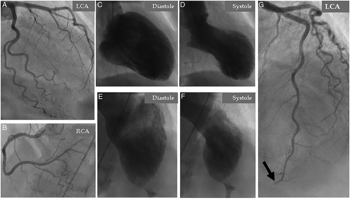
Coronary angiography: (A), left coronary artery (LCA), and (B), right coronary artery (RCA) deemed initially to be normal. Contrast left ventriculography in two projections; right anterior oblique (C and D) and left cranial anterior oblique (E and F) shows circumferential mid‐apical ballooning and normal contractility of the basal segments during systole. Careful review of coronary angiography reveals an abrupt occlusion of the apical segment of the left anterior descending artery (LAD) (G, black arrow)