
FIG 4.
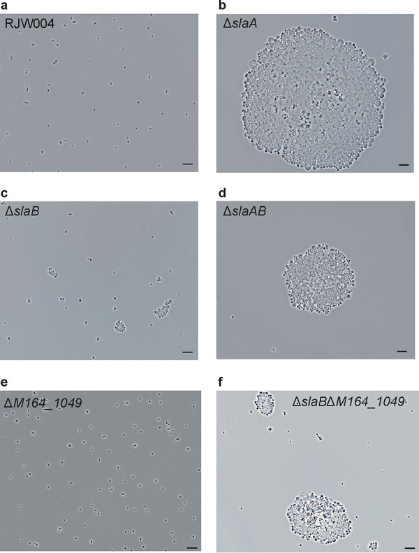
Phenotypic characterization of S-layer gene knockout mutants under a phase-contrast microscope. Representative phase-contrast microscopy images of cells of wild-type RJW004 and mutants are shown, as indicated. Five-microliter cell cultures were spotted on a cleaned microscope slide, covered with a coverslip, and then observed under a microscope. Scale bar, 10 μm.