FIG 2.
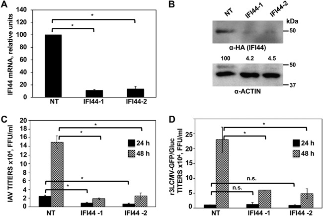
IFI44 silencing negatively affects IAV and LCMV replication. (A) Human A549 cells were transfected with NT or IFI44 siRNAs. At 36 hpt, total RNAs were purified and mRNA levels for IFI44 were analyzed by RT-qPCR. (B) At 36 h after siRNA transfection, cells were transfected for 48 h with the plasmid expressing IFI44 fused to an HA tag. A Western blot analysis using anti-HA antibodies (to detect IFI44; top) and anti-actin antibodies (bottom) was performed. Western blots were quantified by densitometry using ImageJ software. IFI44 protein expression levels in cells silenced with the NT siRNA were assigned a value of 100% for comparisons with the levels of expression in IFI44-silenced cells (numbers below the HA blot). IFI44 expression was normalized to actin expression. Molecular weight markers are indicated (in kilodaltons) on the right. Three different experiments were performed, with similar results. (C and D) At 36 hpt, cells were infected with influenza PR8 virus (C) or r3LCMV-GFP/Gluc (D). Tissue culture supernatants were collected at 24 and 48 hpi and titrated by immunofocus assay (PR8) or fluorescence expression analysis (r3LCMV-GFP/Gluc). Bars represent SDs determined using triplicate wells. Three different experiments were performed, with similar results. *, P < 0.05 (for comparisons between NT- and IFI44-silenced cells at 24 and 48 hpi using Student´s t test). n.s., differences not significant (P > 0.05).

