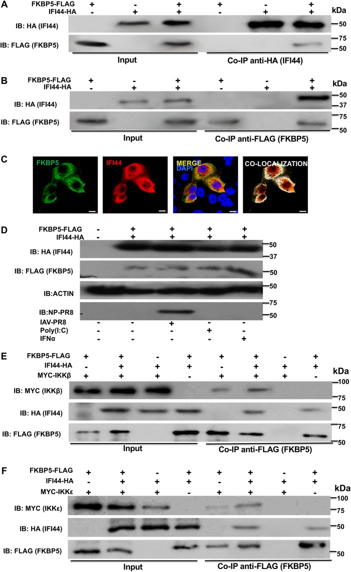
FIG 5.
IFI44 interacts with FKBP5 and does not inhibit binding of FKBP5 to IKKβ or IKKε. (A to C) Human 293T cells were transiently cotransfected with the pCAGGS plasmid encoding IFI44-HA and FKBP5-FLAG, or with empty plasmids, as internal controls. IB, immunoblot. (A and B) Coimmunoprecipitation experiments using anti-HA to pull down IFI44 (A) and anti-FLAG to pull down FKBP5 (B) using affinity columns were performed. Western blotting using antibodies specific for the HA tag (to detect IFI44) or the FLAG tag (to detect FKBP5 protein) was performed to detect protein in the cellular lysates (Input) and after the Co-IP. Molecular weight markers are indicated (in kilodaltons) on the right. Three different experiments were performed, with similar results. (C) At 24 hpi, cells were fixed with paraformaldehyde, FKBP5-FLAG and IFI44-HA were labeled with antibodies specific for the tags (in green and red, respectively), and nuclei were stained with DAPI (in blue). Areas of colocalization of both proteins appear in yellow in the third picture and in white in the fourth picture. Scale bar, 10 μm. (D) Human A549 cells were transiently cotransfected with the pCAGGS plasmid encoding IFI44-HA and FKBP5-FLAG, or with empty plasmids, as internal controls. At 24 hpt, the cells were subjected to mock treatment, transfected with poly(I·C), treated with IFN-α, or infected with IAV for 24 h. Western blotting using antibodies specific for the HA tag (to detect IFI44), the FLAG tag (to detect FKBP5), anti-actin, and IAV anti-NP proteins was performed. Molecular weight markers are indicated (in kilodaltons) on the right. Two different experiments were performed, with similar results. (E and F) Human 293T cells were transiently cotransfected with different combinations of pCAGGS plasmids encoding IFI44-HA, FKBP5-FLAG, and MYC-IKKβ (E) or IFI44-HA, FKBP5-FLAG, and MYC-IKKε (F). Co-IP experiments using an anti-FLAG affinity column (to pull down FKBP5) were performed. Western blotting using antibodies specific for the MYC tag (to detect IKKβ or IKKε), the HA tag (to detect IFI44), and the FLAG tag (to detect FKBP5) was performed to detect protein in the cellular lysates (Input) and after the Co-IP. Molecular weight markers are indicated (in kilodaltons) on the right. Three different experiments were performed, with similar results.

