FIG 6.
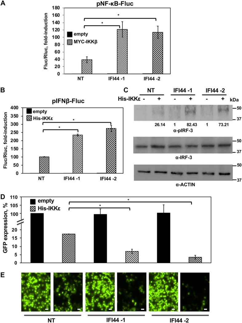
IFI44 negatively affects IKKβ and IKKε activation. (A) IFI44-silenced human 293T cells were transiently cotransfected with a plasmid expressing IKKβ, a plasmid expressing the Fluc reporter gene under the control of NF-κB (pNF-κB-Fluc), and a plasmid constitutively expressing Rluc. (B) IFI44-silenced human 293T cells were transiently cotransfected with a plasmid expressing IKKε together with a plasmid expressing the Fluc reporter gene under the control of IFN-β promoter (pIFNβ-Fluc) and a plasmid constitutively expressing Rluc. (A and B) At 24 hpt, levels of Fluc were determined and normalized to the levels of Rluc. Data represents means and SDs of results from triplicate wells. Experiments were repeated three times with similar results. (C) Cellular lysates from cells analyzed as described for panel B were collected, and protein levels of pIRF-3 and actin were evaluated by Western blotting. Western blots were quantified by densitometry using ImageJ software (v1.46), and the amounts of pIRF-3 were normalized to the amounts of actin (numbers below the pIRF-3 blot). Molecular weight markers are indicated (in kilodaltons) at the right. (D and E) Tissue culture supernatants from cells analyzed as described for panel B were collected and used to treat fresh A549 cells. After 24 h of incubation, cells were infected (MOI of 0.1) with rVSV-GFP. At 24 hpi, GFP expression was quantified in a microplate reader (D) and GFP-infected cells were analyzed by visualizing GFP expression under a fluorescence microscope (E). Experiments were repeated three times with similar results. *, P < 0.05 (using Student’s t test [panels A, B, and D]). Representative images from a microscope using a 20× objective are shown in panel E. Scale bars, 50 μm.

