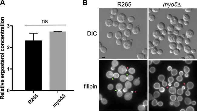
FIG 8.
Ergosterol distribution appears to be altered in the myo5Δ mutant. (A) Ergosterol content is similar between the wild type and myo5Δ mutant. Ergosterol was extracted from cells and quantitated by LCMS. The experiments were repeated three times, and the error bars represent the standard deviations. (B) Filipin staining patterns are different between the wild type and the myo5Δ mutant. Log-phase grown cells were stained with 5 μg/ml filipin. The fluorescent and DIC images were photographed. Red arrowhead, emerging bud; green arrowhead, isthmus. Bar = 2 μm.

