Figure 3.

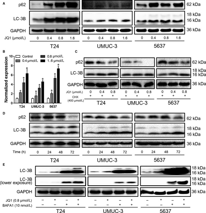
JQ1 induces autophagy flux in BC cells. After 24 h treatment, the expression of p62 and LC‐3B was detected by western blotting analysis (A), the mRNA level of p62 was also determined by RT‐qPCR assay (B). (C) Cells were pretreated with 400 nmol/L cyclohexane (CHX) for 4 h, and then treated with JQ1 in the presence of 400 nmol/L CHX. The expression of p62 and LC‐3B was detected by western blotting analysis. (D) Cells were treated with 0.4 μmol/L JQ1 for different time points, the expression of p62 and LC‐3B was detected by western blotting analysis. (E) Cells were treated with 0.8 μmol/L JQ1 for 12 h, then the culture medium was replaced with fresh medium containing 0.8 μmol/L JQ1 and 10 nmol/L BAFA1. After 12 h, cells were harvested and the expression of LC‐3B was detected by western blotting analysis. *P < 0.05 vs control. Data represent the results of three independent experiments
