Fig. 5.
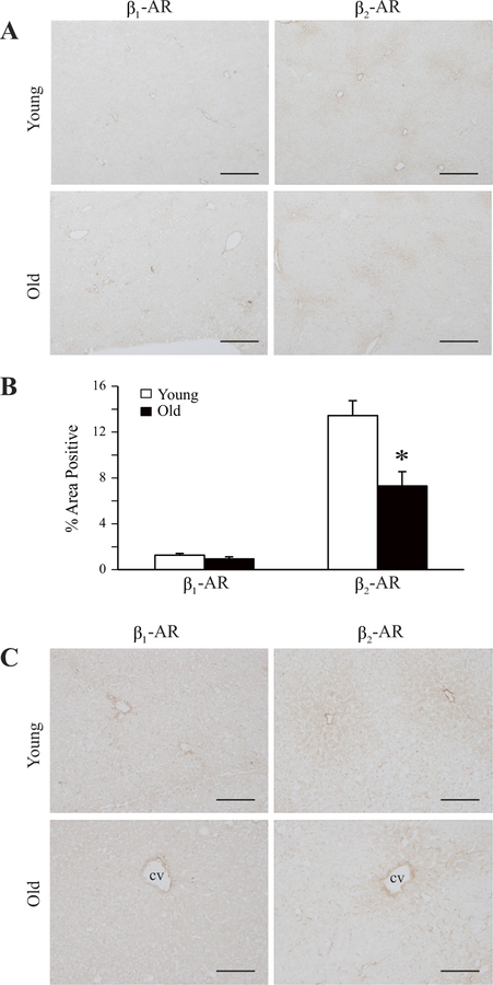
β1- and β2-AR immunoreactivity in liver sections from young and old rats. Frozen liver sections were immunostained for β1-AR and β2-AR. A: representative diaminobenzidine (DAB)-stained liver sections from individual young (6 mo old) and old (24 mo old) rats are shown (×4 objective magnification). Scale bar, 300 µm. B: areas of staining for β1- and β2-AR proteins in young vs. old rat liver sections, as in A, determined by quantitation of DAB staining, are represented as bar graphs. Antibody to β1-AR yielded relatively low signal. Data are expressed as means ± SE from 8 to 9 young and 6 to 8 old rats. *P = 0.002. C: representative liver sections exhibiting greater detail are shown at higher magnification (×12.5 objective). Scale bar, 100 µm. cv, Central vein.

