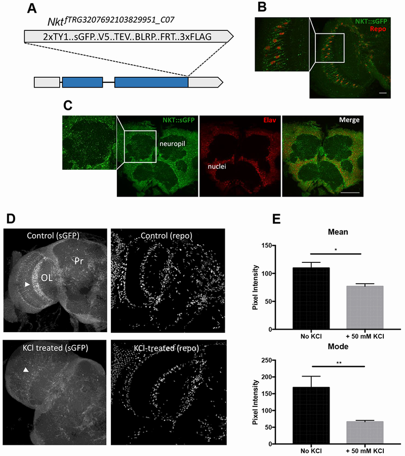
Figure 3. NKT:sGFP is trafficked to glial and neuronal processes and it behaves as a secreted protein.

A) Schematic representation of endogenously tagged Nkt. B) NKT::sGFP expression in optic lobe glial cells of transgenic flies expressing the tagged protein from the endogenous promoter. Repo signal shows the positions of glial cell nuclei. C) Endogenous NKT in adult brain cells as assessed by NKT::sGFP localization. Elav signal shows the positions of neuronal nuclei. D) NKT:sGFP signal within astrocytes of the medulla optic lobe (arrowheads) is decreased in 90 sec by 50 mM KCl treatment. Repo antibody signal is not altered by KCl treatment. E) Mean and modal sGFP pixel intensities (arbitrary units) with and without KCl treatment. n=8 brains for both control and KCl-treated samples. Scale bars are 20 μm for B and 50 μm for C. *, p<0.05; **, p<0.01.
