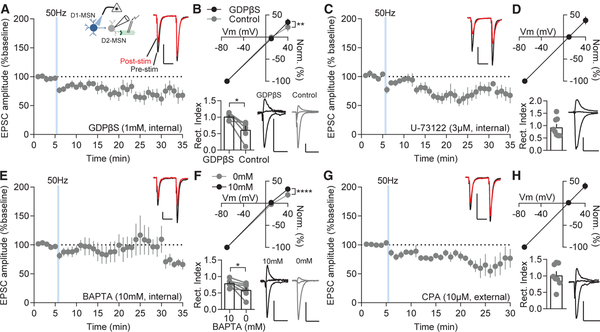
Figure 7. D1-MSN-Stimulation-Induced D2-MSN LLP Requires M1R Signaling.
(A) GDPβS-mediated block of G-protein signaling prevents LLP of NAcC D2-MSNs following D1-MSN 50-Hz stimulation (p < 0.05; n = 8).
(B) Inward rectification of AMPAR currents at +40 mV is blocked by GDPβS compared to D2-MSN pairs (+40 mV: p < 0.01; rectification index p < 0.05; n = 6 and 6).
(C and D) Internal solution with the PLC antagonist U-73122 (C) promotes stimulation-induced depression on D2-MSNs (p < 0.05; n = 9) and (D) prevents inward rectification (p > 0.05; n = 9).
(E and F) Calcium chelation with BAPTA prevents (E) LLP (p > 0.05; n = 9) and (F) inward rectification compared to D2-MSN pairs (+40 mV: p < 0.0001; rectification index p < 0.05; n = 7 and 7).
(G and H) CPA application to deplete internal calcium stores blocks (G) LLP (p > 0.05; n = 8) and (H) inward rectification (p > 0.05; n = 8).
Error bars represent SEM. *p < 0.05, **p < 0.01, ***p < 0.001, ****p < 0.0001. For exact statistics, see Table S1. See also Figure S7.

