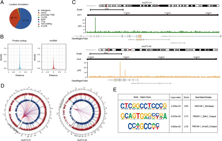
Fig. 4.
Lnc273–31 and lnc273–34 are specifically regulated by p53-R273H in colorectal CSCs. a. Pie chart illustrated the distribution of peak locations (promoter-TSS, TSS, intergenic, exon, intron, 5’UTR, 3’UTR and non-coding) in the genome. The pie chart shows the percentage of peaks in different genomic location. b. The overlap between significant peaks and transcription start sites (TSSs) for various gene classes (mRNAs and lncRNAs) are shown in left (blue) and right (red) of the density plot, respectively. c. Signals of p53 for lnc273–31 (green) and lnc273–34 (yellow), respectively. d. A Circos plot of the top 100 co-expressed genes related to the location of lnc273–31 (left) and lnc273–34 (right), respectively. e. HOMER was utilized to identify motifs. The range of nucleotides around the center of peaks for motif identification was set ±100 bp for establishing the primary and co-enriched motif bound by p53-R273H. Motif identification results were listed in a table including rank, log p value, score, best match/details. Enrichment p-values reported by HOMER was significant (< 0.05)

