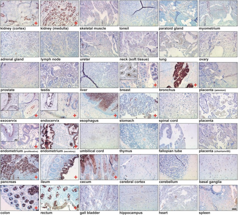
Fig. 1.
CLEC10A staining of various normal human tissues arranged on a tissue microarray. Protein domain histochemistry was performed after complexing of recombinant, myc-tagged CLEC10A with a biotinylated anti-myc antibody conjugated to streptavidin-horseradish peroxidase. 3,3′-diamino-benzidine (DAB) was used as chromogenic substrate and tissues were counterstained with hematoxylin. Tissues stained positive for CLEC10A are marked with “+“. Scale Bar: 100 μm. Inserts with higher magnification of representative tissue areas are given for breast, cervical and endometrium tissues (scale bar: 10 μm)

