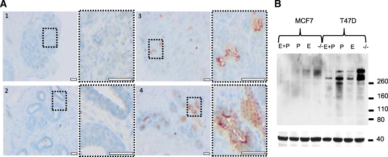
Fig. 2.
Expression of CLEC10A positive glycan structures in normal human breast tissue and breast cancer cell lines is dependent on female sex hormones. a Applying protein domain histochemistry, cryosections of normal human breast tissue obtained during the proliferative phase (#1 + #2) or luteal phase (#3 + #4) of the female hormonal cycle were stained with CLEC10A as described in Fig. 1. Scale bar: 100 μm. Magnifications of the marked areas are given to the right of each overview; scale bar: 100 μm. To determine the phase of the female hormonal cycle, levels of the female sex hormones FSH, LH, 17β-estradiol (E) and progesterone (P) in corresponding serum samples were determined as follows: #1 (age 34 y) FSH: 7.5 IU/ml, LH: 3.9 U/l, E: 57 pg/ml, P: 0.1 ng/ml; #2 (age 43 y) FSH: 6.2 IU/ml, LH: 7.4 U/l, E: 142 pg/ml, P: 0.3 ng/ml; #3 (age 43 y) FSH: 4.1 IU/ml, LH: 5.0 U/l, E: 57 pg/ml, P: 5.3 ng/ml; #4 (age 44 y) FSH: 4.2 IU/ml, LH: 1.2 U/l, E: 116 pg/ml, P: 12.2 ng/ml. Samples were assigned to the luteal phase when serum levels of progesterone were P > 2 ng/ml, FSH < 8 IU/ml and LH in the range between 1 and 11 U/l. b Far Western Blot analysis of total protein extracts (20 μg/lane) from hormone depleted (−/−) MCF7 and T47D cells using recombinant CLEC10A as probe. Cells were cultivated in hormone-depleted medium 4 days before estrogen (E) or progesterone (P) or a combination of both hormones (E + P) was added for 24 h. β-actin served as loading control (lower panel)

