Fig. 6.
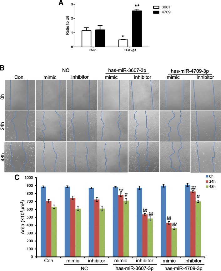
Role of hsa-miR-3607-3p and hsa-miR-4709-3p in TGF-β1-induced migration. a Results of qRT-PCR show the expression of hsa-miR-3607-3p and hsa-miR-4709-3p after stimulation by 5 ng/mL TGF-β1 in HK-2 cells. *p < 0.05, *p < 0.01 versus control group. b Representative images of the wound healing assay. HK-2 cells were treated with 5 ng/mL TGF-β1 for 0, 24, and 48 h. Cells transfected with negative control (NC), hsa-miR-3607-3p and hsa-miR-4709-3p mimics or inhibitor and then treated by TGF-β1. Original magnification: × 100. c Quantitative analysis of the area of cell-free space post-scratch at 0, 24, and 48 h. Data represent mean ± SEM for at least three independent experiments. ***p < 0.01, ***p < 0.001 versus con; ##p < 0.01, ###p < 0.001 versus NC at 24 or 48 h

