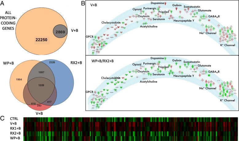
Fig. 2.
JAK/STAT inhibitors reverse BDNF-induced gene expression. a TOP: Venn diagram representation of all protein-coding genes in the rat genome (in accordance with Strand NGS notation) with all differentially expressed genes (DEGs) between primary neurons treated with DMSO+BDNF vs. DMSO+Water. BOTTOM: Venn diagram representation of all DEGs in DMSO+Water vs. DMSO+BDNF (BDNF, Red, 2869), WP1066 + BDNF vs. DMSO+BDNF (WP1066,Orange, 5170 total) and 10 μM Ruxo+BDNF vs. DMSO+BDNF (RX2, Blue, 5393 total genes). b Receptor and Ion Channel expression is altered by BDNF and rescued by JAK inhibition. TOP: List of Ion Channels and Receptor Subunits whose expression is altered by 4-h BDNF treatment. Color represents direction and degree of fold change (Red: up, Green: down) sorted by receptor or channel type. BOTTOM: Receptor and Ion Channel reversal in expression by addition of WP or RX2 (white receptors are not affected by JAK/STAT inhibitors). Response to BDNF in presence of WP was used as the basis for coloring receptors depicted in the diagram. Red: Upregulated, Green: Downregulated. c Heatmap of all DEGs (columns) associated with Epilepsy (IPA) that are affected by exposure to BDNF. DW: DMSO+Water, DB: DMSO+BDNF, RX1: 100 nM Ruxo+BDNF, RX2:10 μM Ruxo+BDNF, WP: 10 μM WP1066 + BDNF. Green: low expression, red: high expression

