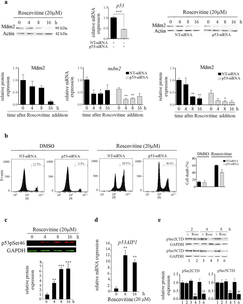
Fig. 4.
Effect of siRNA-mediated downregulation of p53 in ROSC-treated hESCs. a Representative Western blot in H9 cells of Mdm2 at different time points after ROSC addition. Bar graphs show densitometric quantification. Analysis of p53 (upper panel) and mdm2 (lower panel) mRNA expression levels by Real Time RT-PCR in non-targeting (NT) or p53-siRNA transfected hESCs in the presence or absence of ROSC. Representative Western blot in siRNA transfected H9 cells of Mdm2 at different time points after ROSC addition. Bar graphs show densitometric quantification. b H9 hESCs were transfected with NT-siRNA or p53-siRNA and exposed to ROSC (20 μM during 16 h). Representative histograms of PI-stained transfected cells left untreated or exposed to ROSC. Bar graphs show the percentage of cell death. Each bar represents the mean ± SD of three independent experiments. c Time course of p53 phosphorylation at serine 46 upon ROSC treatment was analyzed by Western blotting using anti-phospho-p53Ser46 (p53pSer46) specific antibody. GAPDH served as loading control. d Time-course analyses of p53Ser46 target p53AIP1 mRNA expression levels by Real Time RT-PCR in ROSC-treated or untreated hESCs. Rpl7 expression was used as normalizer. In all cases, a paired Student’s t test was used to test for significant differences between ROSC-treated and untreated samples *P < 0.05, **P < 0.01, ***P < 0.001. e Cells were treated with vehicle (v) (lanes 1–3-5) or 20 μM ROSC (lanes 2–4-6). At the indicated time points, cell lysates were analyzed by immunoblotting using the indicated antibodies. Bar graphs show densitometric quantification

