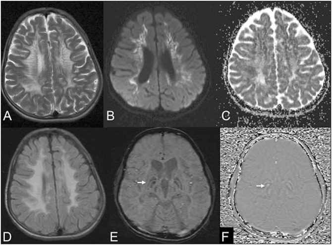
Figure 1.

MRI of a 17-year-old boy with neuropsychiatric manifestations and dystonia. Axial images T2W (A) and FLAIR (D) show confluent periventricular white matter hyperintensities with a classical tigroid appearance in T2. DWI (B) and apparent diffusion coefficient (C) show diffusion restriction in affected white matter. SWI (E) shows prominent susceptibility (arrows) involving the GPi bilaterally with positive phase shift on phase images (F) consistent with exaggerated mineralization
