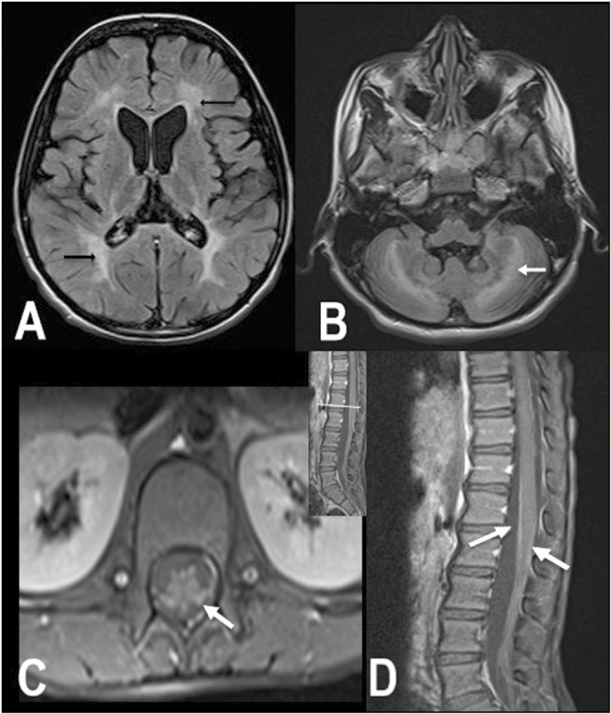
Figure 2.

MRI brain and spine images of P-11. Axial FLAIR shows bilateral confluent symmetric periventricular and deep white matter hyperintensities (arrows) in both the cerebrum (A) and cerebellum (B). Contrast-enhanced fat saturated T1 axial (C) and sagittal (D) images of the lumbar spine show smooth enhancement of the cauda equina nerve roots (arrows)
