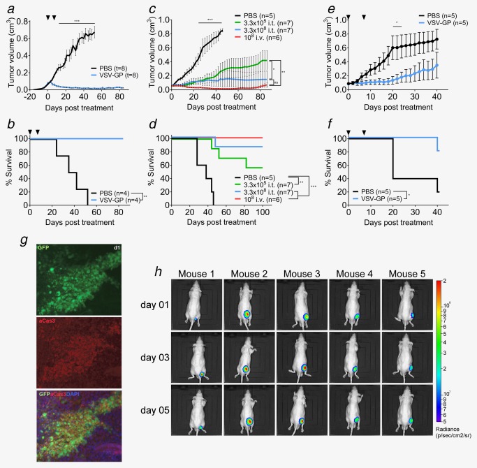
Figure 4.

VSV‐GP shows efficacy in PCa xenograft subcutaneous models. (a,b) 2 × 10⁶ Du145 cells were injected subcutaneously into both flanks of Balb/c‐rag−/−γ−/− mice. Tumors were treated at a size of 0.07 cm3 with intratumoral injection of either 107 pfus VSV‐GP or PBS. Treatment was repeated 7 days later. Mean of tumor volume (a) and Kaplan–Meier survival curve (b) are shown. (c,d) 2 × 10⁶ 22Rv1 cells were injected subcutaneously into the right flanks of nude mice. Tumors were treated at a size of 0.05 cm³ with intratumoral injection of either 3.3 × 10⁵ pfus VSV‐GP, 2.3 × 10⁸ pfus VSV‐GP or PBS or with intravenous injection of 108 pfus VSV‐GP. Mean of tumor volume (c) and Kaplan–Meier survival curve (d) are shown. (e,f) 5 × 10⁶ VCaP cells mixed 1:1 with matrigel were injected subcutaneously into the right flank of nude mice. Tumors were treated at a size of 0.07 cm³ with an intratumoral injection of either 107 pfus VSV‐GP or PBS. Treatment was repeated 7 days later. Mean of tumor volume (e) and Kaplan–Meier survival curve (f) are shown. (g) Subcutaneous 22Rv1 tumors were treated at a size of 0.07 cm³ with an intratumoral injection of 108 pfus VSV‐GP‐GFP. Tumors were isolated at indicated time points and fixed. Cryo‐slices were prepared and probed with fluorescently labelled antibodies recognizing activated caspase 3. Antibody signal and viral GFP signal were analyzed under the fluorescence microscope. (h) Subcutaneous 22Rv1 tumors treated at a size of 0.07 cm³ with an intratumoral injection of 108 pfus VSV‐GP‐Luc. Representative BLI pictures of treated mice are shown.
