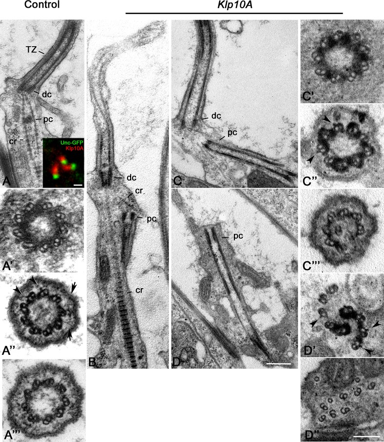
FIGURE 6.
Ultrastructural defects in Johnston’s organs of Klp10A mutant flies. Control Johnston’s organs: Longitudinal section (A); Cross section at the level of the distal centriole (A’), the transition zone (A”), the proximal region of the axoneme (A”’). Klp10A is localized just above the Unc-GFP dot in sensory cilia (inset A). Klp10A Johnston’s organs: longitudinal sections of the whole ciliary complex (B), proximal and distal centrioles (C) and proximal centriole alone (D); cross section through the proximal centriole (C’), the transition zone (C”) and the proximal region of the axoneme (C”’); cross section throughout the intermediate (D’) and the distal (D”) regions of the proximal centriole. DC, distal centriole; PC, proximal centriole; CR, ciliary rootlet; TZ, transition zone; arrowheads, lateral projections emerging from the A-tubule; arrows, Y-links. Bars: 0.4 mm (A–D); 1 mm (inset A); 50 nm (A’–A”’C’–C”’D’,D”).

