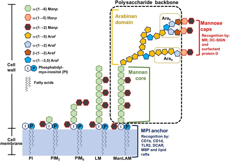
Figure 1.
ManLAM structure. ManLAM biosynthesis follows a pathway from phosphatidyl-myo-inositol (PI)→PIM→LM→LAM→ManLAM. ManLAM contains three domains: an MPI anchor, a polysaccharide backbone and mannose caps. The MPI anchor comprises a PI unit with Manp units. PI acts as an anchor inserted into the cell membrane. The MPI anchor is recognized by CD1b, CD1d, TLR2, DCAR, MBP and lactosylceramide enriched lipid rafts. The polysaccharide backbone includes a mannan core and an arabinan domain. In the mannan backbone of LAM/ManLAM, PIM2 is linked to another 17–19 residues of Manp. The arabinan core consists of a branched linear α (1→5) linked Araf. Mature LAM/ManLAM is further linked via an arabinan domain made up of approximately 70 Araf residues. Two arrangements or motifs can be found at the non-reducing end: a branched hexaarabinofuranoside (Ara6) and a linear tetraarabinofuranoside (Ara4). The mannose caps consist of one to three Manp residues linked to the terminal β-linked Araf unit. The mannose caps are recognized by MR, DC-SIGN and surfactant protein D.

