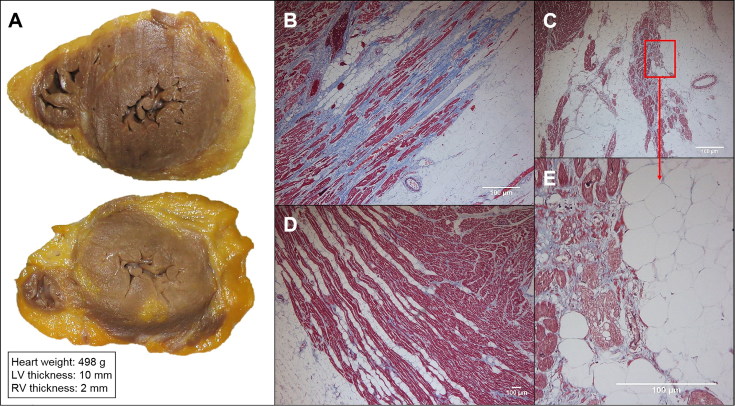
Figure 2.
Macroscopic and histological study of proband’s heart. (A) Cross section of the heart near apex and the immediately above segment. Marked epicardial adipose infiltration with a thick subepicardial band at the left ventricle (LV). Microscopic images of the posterior (B) and lateral (C) wall of the LV that show subepicardial fibrofatty infiltration with different amounts of fibrosis. (D) Right ventricle (RV) showing mild lipomatosis in the external third myocardium, without myocardial degeneration or replacement fibrosis. (E) Detail of the myocyte degeneration.

