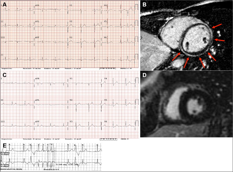
Figure 3.
Images from the cardiological work-up in living mutation carriers. (A) Resting electrocardiogram (ECG) from the proband’s distant relative in whom the genetic study was performed, with a left dominant arrhythmogenic cardiomyopathy (LDAC) phenotype. (B) Subepicardial extensive late gadolinium enhancement in the same individual as in (A). (C) Resting ECG from the proband’s sister with palpitations. (D) Normal cardiac magnetic resonance imaging without late gadolinium enhancement in the same patient as in (C). (E) A run of non-sustained ventricular tachycardia recorded at the holter ECG in the same patient as in (C).

