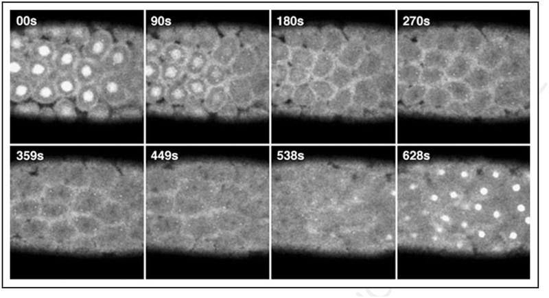
Figure 3.
Panel of time-lapse confocal images of GFP fluorescence taken at the cortical surface of a homozygous GFP-Zw3 syncytial Drosophila embryo. Note that although centrosomes tend to move out of the plane of focus during time-lapse imaging (e.g., panels 359 s to 628 s), we find GFP-Zw3 localized to centrosomes throughout rounds of syncytial mitosis. Shown are selected frames at the times indicated after centrosome separation during prophase († = 00 s) and progress through anaphase (~† = 449 s later). Chromosomes, which do not fluoresce and appear as dark shadows, can be seen in roughly metaphase configuration in panels 270 to 359s. S-phase of the subsequent round of division is shown in the last panel († = 628 s). At room temperature, rounds of division in both wt and GFP-Zw3 embryos occurred approximately every 10.5 min. Time units are in seconds (s) and scale bar is 50 μm.

