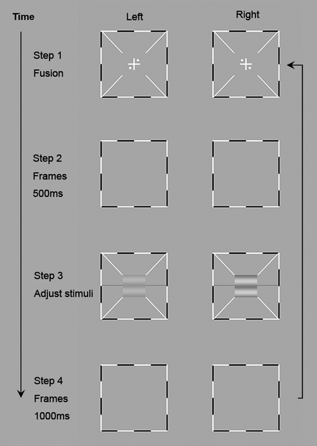
Figure 2.
Binocular phase combination procedure22. The left column shows the stimuli in the left eye and the right column shows the stimuli in the right eye. In step 1, subjects adjusted the two frames to fuse the images displayed to the left and right eyes. After perceiving one cross with four dots in the four quadrants, they could press a key to start the trial. A blank frame appeared for 500ms in Step 2. In Step 3 horizontal sine-wave gratings were presented to the two eyes and subjects adjusted the reference line to indicate the “darkest” part of the cyclopean grating. A blank frame was displayed for 1000 ms after the subject finished this trial (Step 4).

